Dental X-rays
A typical case, in 6 steps over 1 visit. Typically dental X-rays use very low radiation, and they are generally considered safe during pregnancy. Every step names its source.
Not yet reviewed by a licensed clinician. Every step names its source.
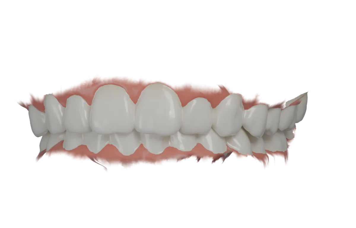
Dental X-rays · Visit 1
Deciding which images you need
Your dentist decides whether you need X-rays and which kind, based on your current oral health, age, risk for disease and any symptoms. There is no one-size-fits-all schedule, and you can talk it over with your dentist.
Source: ada.org
Steps in a typical case
Deciding which images you need
Your dentist decides whether you need X-rays and which kind, based on your current oral health, age, risk for disease and any symptoms. There is no one-size-fits-all schedule, and you can talk it over with your dentist.
Visit 1 · Source 1
Getting into position
You sit or stand in front of the machine while the technician positions the film or sensor and takes the image. Stay as still as possible.
Visit 1 · Source 2
Images from inside the mouth
For these, the film or sensor goes inside your mouth. Bitewing images show cavities between teeth and below the gumline. Periapical images show gum disease, bone loss and cavities near the tooth roots.
Visit 1 · Source 2
Panoramic image
A panoramic X-ray is taken with the sensor outside your mouth. It shows your entire mouth, including your teeth, jaws, nerves, sinuses and bone.
Visit 1 · Source 2
Looking at the images
Your dentist uses the images to look for cavities, decay under existing fillings, bone loss, impacted teeth and infections. They also help plan treatments like implants, braces or dentures, and track healing.
Visit 1 · Source 2
Keeping exposure low
Dental X-rays use a very low amount of radiation. Digital X-rays use 80% to 90% less radiation than film. Dentists also limit the X-ray beam to the area that needs to be examined and try to use images from earlier exams.
Visit 1 · Source 2
Typical trade-offs
| Visits | Typically the X-rays are taken during the dental visit. Source 2 |
|---|---|
| Time to final result | Typically dental X-rays use very low radiation, and they are generally considered safe during pregnancy. Source 2 |
| How long it lasts | Typically people with healthy teeth and gums need new X-rays only every six to 18 months, and more often if they have gum disease or frequent cavities. Source 2 |
| Upkeep | Typical upkeep: talk with the dentist about whether you need X-rays, because how often depends on oral health, age, risk for disease and any symptoms. Source 1 |
Figures for a typical case from the sources below. A dentist can say how a given case differs.
Questions to ask your dentist
- The typical case takes one visit. Would mine, and how long would it take?
- What are my other options, including waiting, and what happens if I wait?
- How long does it usually last in a case like mine, and what upkeep will it need?
- Will it hurt, and what can I eat or do while it heals?
- What will it cost me, and will my insurance cover it? Ask which billing codes apply (the typical case uses D0210, D0274, D0330).
- How many of these have you done, and who would do mine?
Sources
- X-rays and radiographs. American Dental Association. www.ada.org/resources/ada-library/oral-health-topics/x-rays-radiographs. Accessed November 14, 2026.
- Dental X-rays. Cleveland Clinic. my.clevelandclinic.org/health/diagnostics/11199-dental-x-rays. Accessed November 14, 2026.
Osler Press procedure library, version 2026.11.0. Billing codes for the typical case: D0210, D0274, D0330.